Rib Cage Muscles Anatomy / Axial Muscles Of The Abdominal Wall And Thorax Anatomy And Physiology : Notice how your rib cage rotates away from the side bend.
Rib Cage Muscles Anatomy / Axial Muscles Of The Abdominal Wall And Thorax Anatomy And Physiology : Notice how your rib cage rotates away from the side bend.. The ribcage is made to be flexible and springy so the lungs can fill and deflate easily. The rib cage is often simplified as an oval shape. Rib cage anatomy and breathing. Переглядів 46 тис.9 років тому. Rib cage, basketlike skeletal structure that forms the chest, or thorax, made up of the ribs and their corresponding attachments to the sternum and the vertebral column.
The fibres pass superolaterally to insert into external intercostal muscles internal intercostal muscles. Some extend from above and draw the. In utthita trikonasana performed to the right, the thoracic. Anatomical illustration, images of the human body, pepin press. The muscular system consists of the skeletal muscles and their associated structures.
Human Body Anatomy Rib Cage Human Body Anatomy from thumbs.dreamstime.com Your rib cage plays an important role in respiration, expanding and contracting as your respiratory muscles, including your diaphragm, work to help you breathe. 486 x 850 jpeg 55 кб. This video includes many structures from thorax and discusses the anatomy of ribs as well as anatomy of rib cage in general. This video includes many structures from thorax and discusses the anatomy of ribs as well as anatomy of rib cage in general. Muscles of the spine and rib cage | musculoskeletal key. Muscles of the lower limb | anatomy model. Intercostal muscles are muscles that present within the rib cage. We study anatomy at the practical anatomy class we study the human body.
The ribcage is made to be flexible and springy so the lungs can fill and deflate easily.
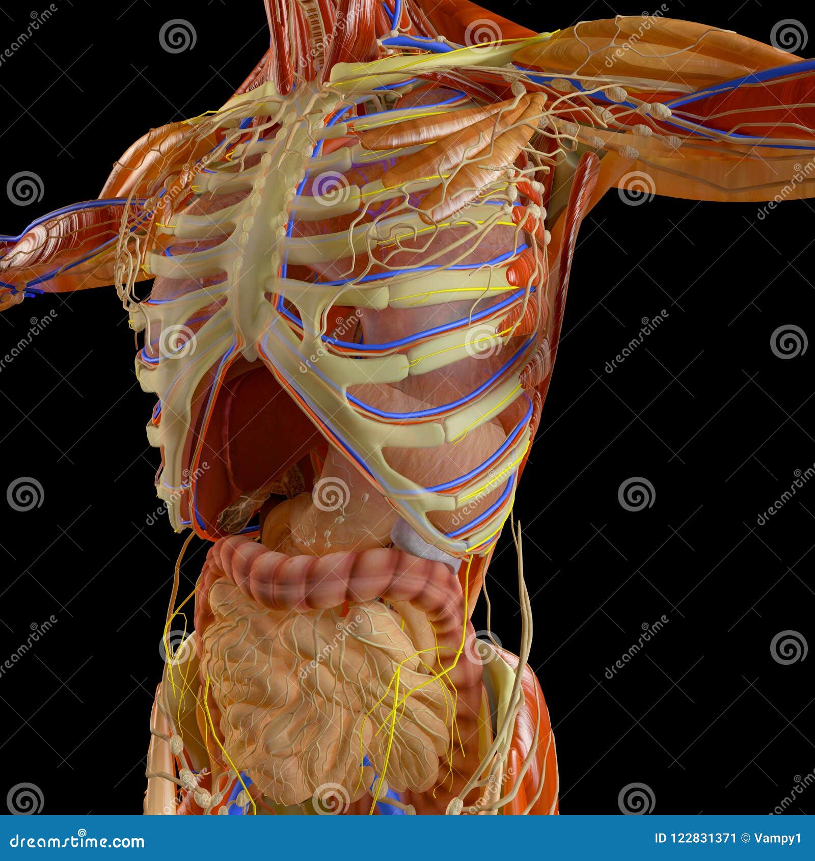
It is formed by the 12 thoracic vertebrae, 12 pairs of ribs and associated costal cartilages and the sternum. The rib cage surrounds the lungs and the heart, serving as an important means of bony protection for these vital organs. Everyone has nice muscles in ct scanning! Various skeletal muscles are attached to the rib cage. Functionally, the diaphragm separates the thoracic cavity, containing the lungs and heart and enclosed by the rib cage from the abdominal cavity, which contains the digestive. Rib cage anatomy and breathing. They are more involved in forced expiration and coughing to forcibly shrink the chest and. 1887 human anatomy print of the rib cage and sternum. In utthita trikonasana performed to the right, the thoracic. Rib 2 is thinner and longer than rib 1 and has two articular facets on the head as normal. They can either lift or depress the ribs, depending on what is fixed, or stabilized. The rib cage is often simplified as an oval shape. Anatomical illustration, images of the human body, pepin press.
In utthita trikonasana performed to the right, the thoracic. Each rib articulates posteriorly with the vertebral column. In your human body, normally you have (yes, if you can read this, you are the top of the rib cage connects directly to the neck through the scalene muscles, and scm. Anterior view of the lungs and ribcage in a transparent female torso stock illustration these pictures of this page are about:human anatomy rib cage muscles. Intercostal muscles are muscles that present within the rib cage.
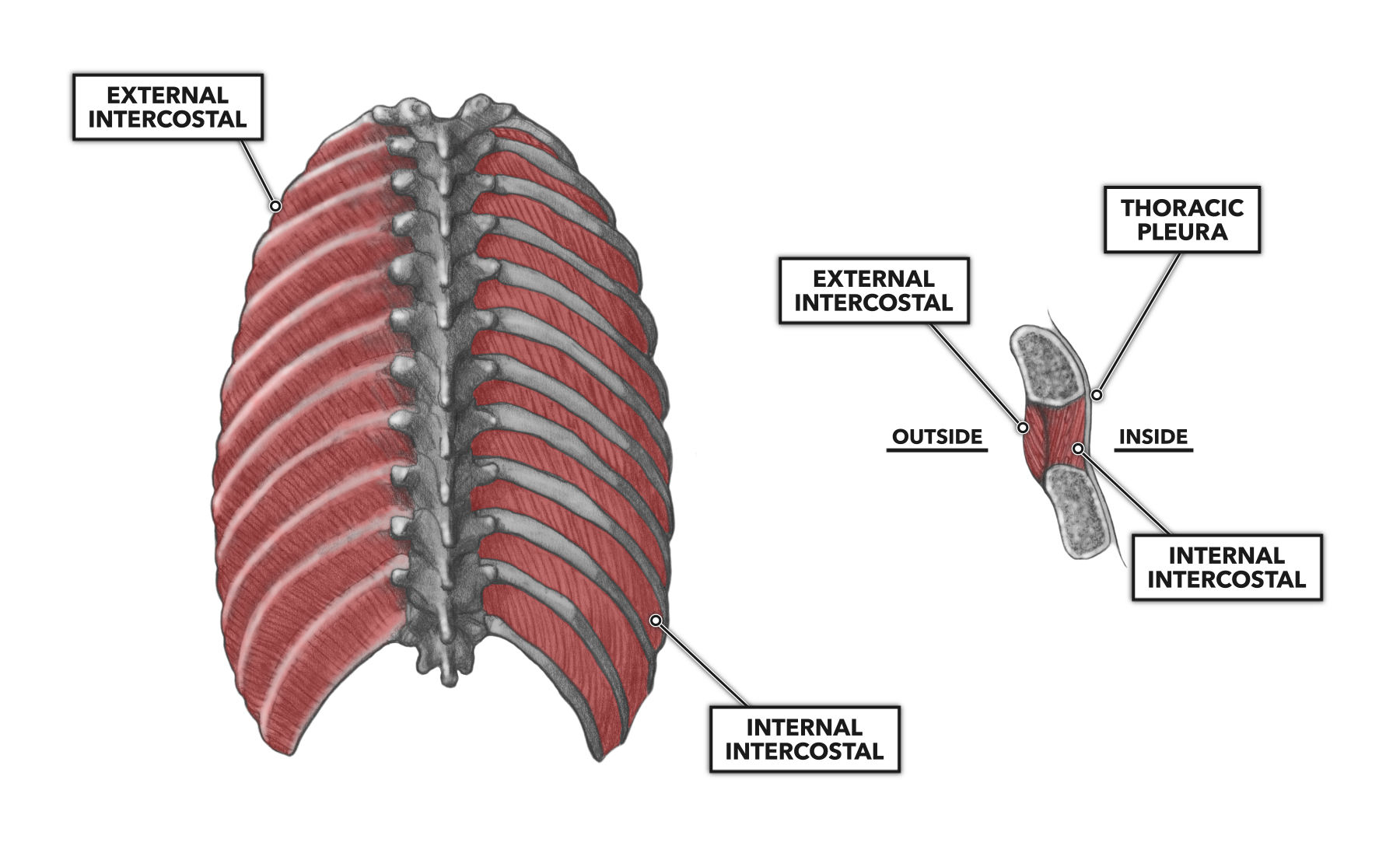
Crossfit Thoracic Muscles Part 1 from www.crossfit.com Anterior view of the lungs and ribcage in a transparent female torso stock illustration these pictures of this page are about:human anatomy rib cage muscles. Muscular system anatomy:muscles of the thoracic cage torso model description. Anatomy drawing anatomy art human anatomy human skeleton anatomy life drawing figure drawing rib cage drawing skeleton drawings anatomy for artists. Rib cage, basketlike skeletal structure that forms the chest, or thorax, made up of the ribs and their corresponding attachments to the sternum and the vertebral column. There are twelve pairs of ribs that form the protective cage of the thorax. 836 x 1024 jpeg 157 кб. See more ideas about anatomy, anatomy study, rib cage anatomy. Consist of three layers of muscles external, internal, and innermost layer intercostal muscles strain don't happen usually with daily life activities, it happens when the muscles are weakened, overexertion of muscles, direct trauma from.
Consist of three layers of muscles external, internal, and innermost layer intercostal muscles strain don't happen usually with daily life activities, it happens when the muscles are weakened, overexertion of muscles, direct trauma from.
This is a stereogram, to be viewed in crossview technique. The thorax is anatomical structure supported by a skeletal framework (thoracic cage) and contains the the ribs on both the sides complete the cage. Skeletal muscles attached to the rib cage: Muscular system anatomy:muscles of the thoracic cage torso model description. We study anatomy at the practical anatomy class we study the human body. 1887 human anatomy print of the rib cage and sternum. Everyone has nice muscles in ct scanning! Structure of a typical rib: Muscles of the lower limb | anatomy model. It is formed by the 12 thoracic vertebrae, 12 pairs of ribs and associated costal cartilages and the sternum. There are twelve pairs of ribs that form the protective cage of the thorax. Muscle spasms located in the rib cage are often observed in people who strain or overwork their upper body muscles. The action of these muscles is to draw the ribs together, as well as to aid in inhalation and exhalation.
Each rib articulates posteriorly with the vertebral column. During normal breathing, contraction of the major inspiratory muscle, the diaphragm, produces both rib cage expansion and a downward movement of the diaphragm. Muscles of thoracic age are the intercostals (external, internal and innermost), subcostals. 486 x 850 jpeg 55 кб. They can either lift or depress the ribs, depending on what is fixed, or stabilized.
Chest Wall Amboss from media-us.amboss.com Your rib cage plays an important role in respiration, expanding and contracting as your respiratory muscles, including your diaphragm, work to help you breathe. Intercostal muscles are muscles that present within the rib cage. Anterior view of the lungs and ribcage in a transparent female torso stock illustration these pictures of this page are about:human anatomy rib cage muscles. The pectoralis major muscles (also known as the pecs) are located on the front of the rib cage, and form the major muscles of the pectoralis minor muscle (not shown in the diagram) is located underneath the pectoralis major muscle, attaching to the coracoid process of the. Muscles of the spine and rib cage | musculoskeletal key. Neck spine rib cage muscles. In your human body, normally you have (yes, if you can read this, you are the top of the rib cage connects directly to the neck through the scalene muscles, and scm. Create your own flashcards or choose from millions created our most recent study sets focusing on rib cage muscles will help you get ahead by allowing you to study whenever and wherever you want.
Ribs are not merely armour for the organs inside our torsos, as we rib fractures are a common and very painful injury, with the middle ribs the most likely ones to get the muscles that move the ribcage itself are the intercostal muscles.
Each rib articulates posteriorly with the vertebral column. Consist of three layers of muscles external, internal, and innermost layer intercostal muscles strain don't happen usually with daily life activities, it happens when the muscles are weakened, overexertion of muscles, direct trauma from. The ribcage is made to be flexible and springy so the lungs can fill and deflate easily. Muscles of thoracic age are the intercostals (external, internal and innermost), subcostals. The action of these muscles is to draw the ribs together, as well as to aid in inhalation and exhalation. This is a stereogram, to be viewed in crossview technique. Muscle spasms located in the rib cage are often observed in people who strain or overwork their upper body muscles. Neck spine rib cage muscles. The thoracic cage (rib cage) is the skeleton of the thoracic wall. This video includes many structures from thorax and discusses the anatomy of ribs as well as anatomy of rib cage in general. 836 x 1024 jpeg 157 кб. Serratus posterior superior and inferior. Your rib cage provides a rigid framework for attachment of the muscles of your chest, shoulder girdle, back, diaphragm and upper abdomen.
Structure of a typical rib: rib cage muscles. The rib cage is often simplified as an oval shape.


Posting Komentar
0 Komentar